Synthesis of 0D lead-free perovskite nanocrystals
0.32 × 1.5 mmol Cs2CO3, 0.35 mmol metal acetate, 15 ml octadecene (ODE), 5 ml oleic acid (OA), and 3 ml oleylamine (OLA) were added in a 50 ml three-neck round-bottom (RB) flask.
The reaction mixture was degassed under a vacuum for 30 minutes at room temperature. Subsequently, the temperature of the reaction mixture was increased to 120°C and vacuumed for another 1 hour to dissolve the Cs2CO3 and terbium acetate.
The RB was filled with N2 gas. The reaction mixture was heated to 220°C for 10 minutes. At that temperature, a solution mixture of 400 μL benzoyl chloride and 500 μL ODE was briskly injected into the RB. Immediately (after 15 seconds) after the injection, the vial was cooled down by an ice-water bath. Then, 2 ml of hexane was added to the solution. The reaction mixture was centrifuged at 8000 rpm for 5 minutes, and the supernatant was decanted.
The precipitated nanoparticles were redispersed in 5 ml of hexane and centrifuged at 4000 rpm for 5 minutes. The supernatant was collected and precipitated by adding 5 ml ethyl acetate and centrifuged at 8000 rpm for 5 minutes. The precipitated nanoparticles were redispersed in 5 ml hexane and centrifuged at 5000 rpm for 5 minutes.
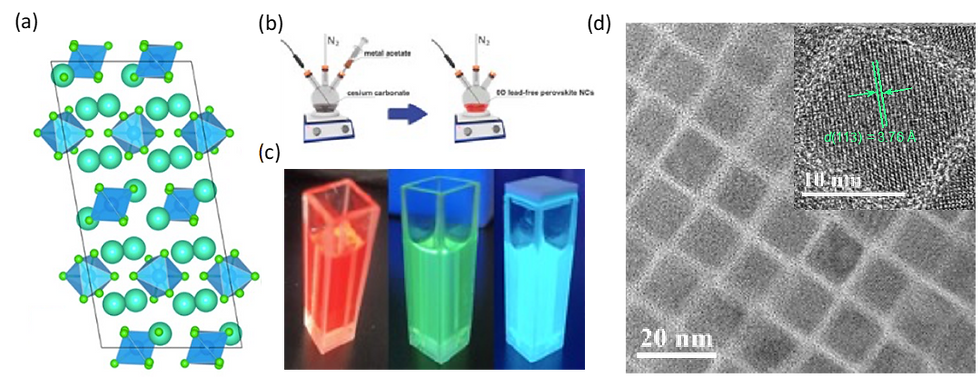
Figure. 1(a) The crystal structure of 0D lead-free perovskite nanocrystals. (b) Schematic of hot-injection method for 0D lead-free perovskite nanocrystal solution. (c) different color images of 0D lead-free perovskite solution under the UV lamp. (d) TEM image of 0D lead-free perovskite nanocrystals
Characterization
The PL and PLE spectra were measured by Hitachi F-7000. The absorbance and XRD spectra of the perovskite films were measured using Cary 5000 UV-Vis-NIR spectrophotometer and an XRD (Miniflex 600) using a diffracted beam monochromator set for Cu-Kα radiation (λ = 1.54056 Å). The 2θ scan range was 10o–60o with a step size of 0.01o, respectively. The TRPL of the samples were measured using a HORIBA spectrophotofluorometer with TCSPC with pulsed nano LED 316 nm (±10 nm). The absolute PLQY of the samples were measured using a HORIBA FluoroMax Plus spectrofluorometer paired with K-sphere petite internal, directly coupled with Integrating Sphere.